Le Dr Sebastian Brandner, MD, neuropathologiste de renom, explique comment les diagnostics moléculaires transforment la prise en charge des tumeurs cérébrales. Il détaille le rôle clé du test de mutation IDH dans les gliomes et les astrocytomes, tout en soulignant les cas où l'histologie classique suffit encore au diagnostic des méningiomes.
More in All


Diagnostic moléculaire en pathologie des tumeurs cérébrales : du méningiome au glioblastome
Aller à la section
- Histologie traditionnelle dans le diagnostic des tumeurs cérébrales
- Diagnostic simple des méningiomes bénins
- Tests moléculaires pour les gliomes et mutations IDH
- Biomarqueurs clés dans le diagnostic des astrocytomes
- Facteurs pronostiques au-delà du type tumoral
- L'avenir du diagnostic des tumeurs cérébrales
- Transcript intégral
Histologie traditionnelle dans le diagnostic des tumeurs cérébrales
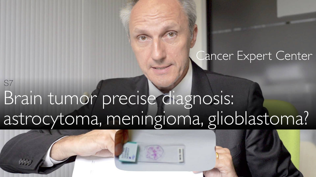
Le Dr Sebastian Brandner souligne que l’histologie de base demeure le fondement de la pathologie des tumeurs cérébrales. La lame colorée à l’H&E (hématoxyline-éosine) fournit des informations initiales essentielles : les échantillons teints en rose révèlent l’architecture tumorale au microscope. Cette méthode traditionnelle continue de guider la démarche diagnostique dans de nombreux cas.
Diagnostic simple des méningiomes bénins
Les méningiomes, issus des enveloppes cérébrales, ne nécessitent souvent aucun test moléculaire, comme l’explique le Dr Sebastian Brandner. Ces tumeurs typiquement bénignes peuvent être diagnostiquées définitivement par la seule histologie standard. Le neuropathologiste note que la localisation tumorale – en particulier au niveau de la base du crâne, difficile d’accès – influence souvent le pronostic davantage que les facteurs moléculaires. Les méningiomes de surface présentent généralement d’excellents résultats après une exérèse chirurgicale complète.
Tests moléculaires pour les gliomes et mutations IDH
Pour les gliomes, surtout chez les patients jeunes, le Dr Sebastian Brandner souligne l’importance cruciale du test de mutation IDH. Un anticorps spécialisé, développé au Centre allemand de recherche sur le cancer, détecte 90 % des mutations IDH1 ainsi que les rares variants IDH2. Cette méthode immunohistochimique, économique et rapide, fournit des résultats fiables qui influencent significativement les décisions thérapeutiques et l’évaluation pronostique.
Biomarqueurs clés dans le diagnostic des astrocytomes
Le Dr Brandner décrit une approche à deux anticorps pour le diagnostic des astrocytomes. Le test de mutation IDH est combiné à l’analyse de la perte protéique nucléaire : lorsque le « point noir » caractéristique disparaît des noyaux cellulaires, cela indique fortement un astrocytome. Cette signature moléculaire aide à différencier les astrocytomes des autres sous-types de gliome et oriente les stratégies thérapeutiques.
Facteurs pronostiques au-delà du type tumoral
Bien que les diagnostics moléculaires fournissent des informations essentielles, le Dr Brandner souligne que la localisation tumorale et l’accessibilité chirurgicale restent des facteurs pronostiques déterminants. Même des tumeurs bénignes situées dans des zones anatomiques difficiles peuvent avoir un pronostic moins favorable que des tumeurs plus agressives mais accessibles. Le neuropathologiste insiste sur la nécessité d’une évaluation complète intégrant données histologiques, moléculaires et cliniques.
L’avenir du diagnostic des tumeurs cérébrales
Le Dr Sebastian Brandner anticipe une avancée continue des techniques de pathologie moléculaire. Le succès du test de mutation IDH montre comment les biomarqueurs ciblés peuvent rationaliser le diagnostic tout en améliorant sa précision. À mesure que la recherche identifie davantage de signatures moléculaires tumorospécifiques, les laboratoires de pathologie combineront de plus en plus méthodes traditionnelles et moléculaires pour optimiser la prise en charge des patients.
Transcript intégral
Dr. Anton Titov, MD: Autrefois, les tumeurs cérébrales étaient diagnostiquées de manière relativement rudimentaire. La coloration de base reste la première étape de l’analyse anatomopathologique. Mais on dispose désormais de davantage d’outils moléculaires et d’analyses des mutations. Cela devient très important pour le traitement et le pronostic des tumeurs cérébrales.
Dr. Anton Titov, MD: Quelle est l’importance des diagnostics moléculaires pour le diagnostic et le traitement des tumeurs cérébrales ?
Dr. Sebastian Brandner, MD: Vous avez tout à fait raison : la base du diagnostic anatomopathologique reste la première lame histologique. C’est une lame colorée en rose, d’environ 1 par 3 pouces. Parfois, on distingue au centre de petites taches roses : ce sont les spécimens de tissu tumoral cérébral.
Dr. Sebastian Brandner, MD: Nous plaçons d’abord cette lame sous le microscope. C’est la première étape du diagnostic.
Dr. Anton Titov, MD: Que fait-on ensuite ?
Dr. Sebastian Brandner, MD: Pour certaines tumeurs, comme le méningiome bénin – qui se développe à partir des enveloppes cérébrales –, le diagnostic peut être posé sur cette seule lame. C’est rapide, économique et très informatif.
Dr. Sebastian Brandner, MD: Aucun test moléculaire supplémentaire n’est nécessaire. D’autres facteurs, comme la localisation de la tumeur – par exemple à la base du crâne, difficile d’accès –, influencent le pronostic. Une tumeur accessible en surface peut être retirée et ne récidive généralement pas. Si un méningiome récidive, une seconde exérèse est souvent possible.
Dr. Sebastian Brandner, MD: Les gliomes aussi peuvent souvent être diagnostiqués sur une lame H&E, notamment les glioblastomes. Mais chez les patients plus jeunes, certains gliomes présentent des mutations IDH.
Dr. Sebastian Brandner, MD: Ces mutations peuvent être détectées à l’aide d’un anticorps développé à Heidelberg, au Centre allemand de recherche sur le cancer. Cet anticorps, économique et rapide, détecte 90 % des mutations IDH (IDH1 dans 95 % des cas, et les rares IDH2).
Dr. Sebastian Brandner, MD: On combine cet anticorps avec un autre qui détecte une protéine nucléaire normalement présente dans le noyau. Dans certains astrocytomes, cette protéine est perdue – le « point noir » disparaît du noyau. Cette perte est caractéristique et quasi diagnostique de l’astrocytome.
Dr. Anton Titov, MD: La mutation IDH est également présente dans l’astrocytome. L’expert explique ainsi le diagnostic moléculaire précis des tumeurs cérébrales : méningiome bénin, astrocytome plus agressif et glioblastome multiforme (GBM).